


For Retinal Physician's Surgical Pearls video series, Melissa Yuan, MD, demonstrates an internal limiting membrane (ILM) peel in a case of macular hole. Transcript of the narration follows below:
This is Melissa Yuan, retina fellow at Stanford, in conjunction with Dr. Ehsan Rahimy, a retina specialist at Palo Alto Medical Foundation and Stanford. This video demonstrates an ILM peel for a macular hole.
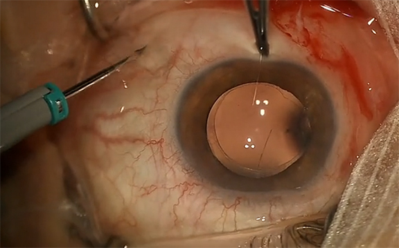
This patient with a history of vitrectomy for retinal detachment now presents with a macular hole that did not improve with drops. He elected to proceed with surgery.
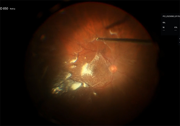
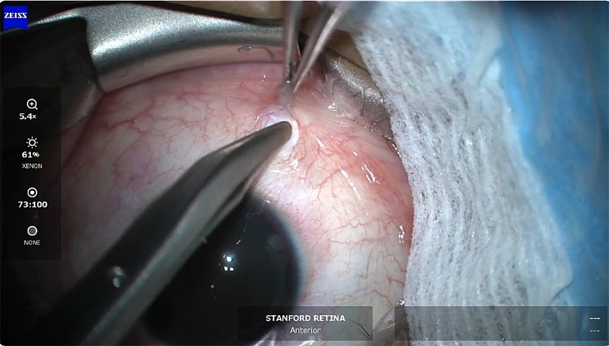
Dilute ICG dye is introduced into the posterior pole. A drop-on contact lens viewing system is placed and the 25-gauge ILM forceps are used to peel the small areas of overlying epiretinal membrane as well as the underlying internal limiting membrane in a circumferential fashion to the hole. Here we are using a pinch and peel technique.
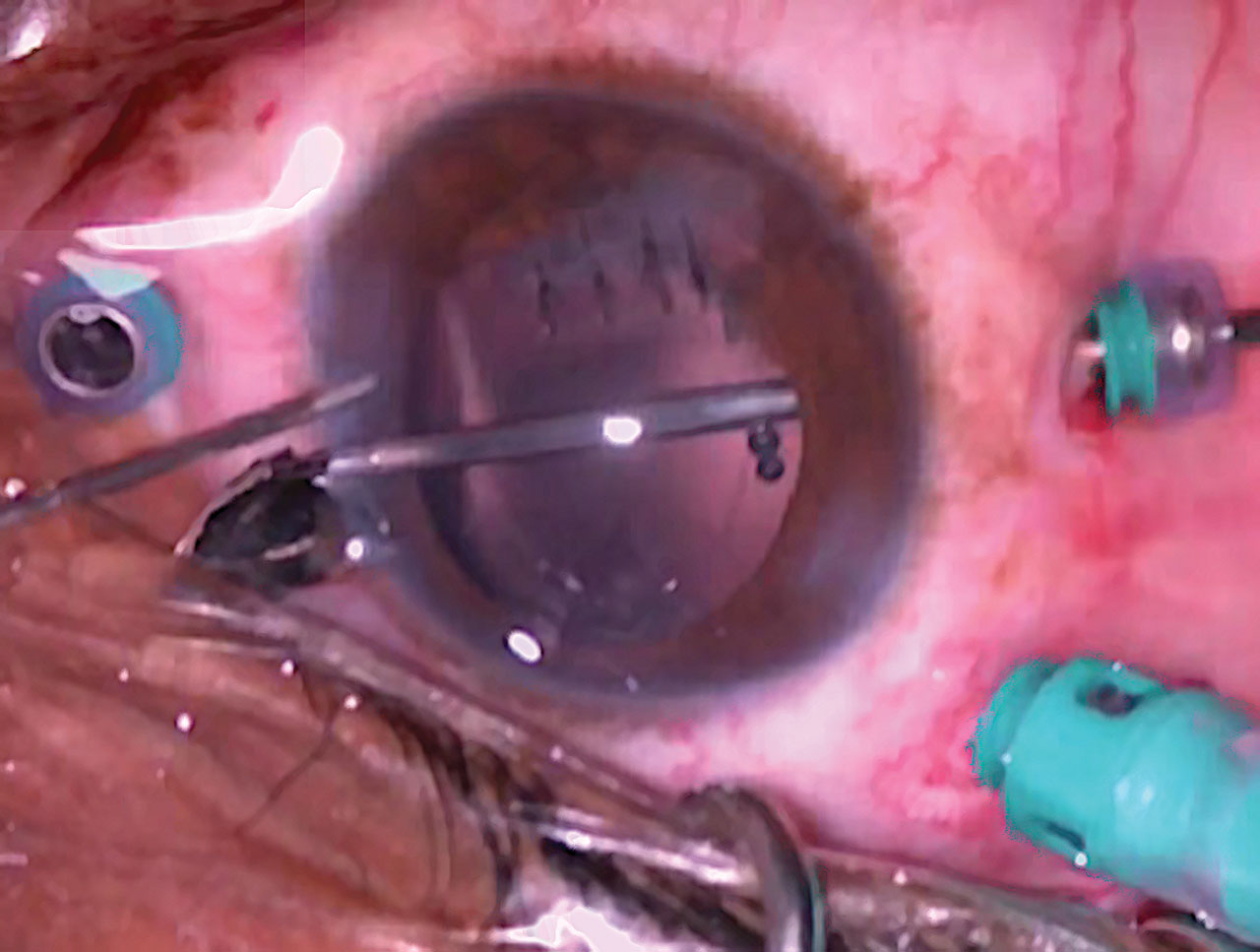
Now we switch to use the cutter on aspiration mode. Using the vitrectomy probe on aspiration only, the ILM flap is then peeled further. The amputated pieces are aspirated.
We switch back to the 25-gauge ILM forceps to complete peeling the ILM to release all of the traction on the fovea.
Fluid air exchange is performed, and the eye is field with 16% C3F8. At postop week 1, the macular hole is closed. RP


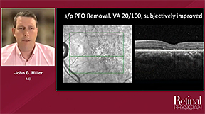
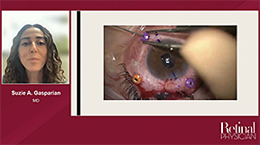
Other Videos From Series